Introduction
Overview of Anatomy and Physiology
Learning Objectives
By the end of this section, you will be able to:
- Compare and contrast anatomy and physiology, including their specializations and methods of study
- Discuss the fundamental relationship between anatomy and physiology
Human anatomy is the scientific study of the body’s structures. Some of these structures are very small and can only be observed and analyzed with the assistance of a microscope. Other larger structures can readily be seen, manipulated, measured, and weighed. The word “anatomy” comes from a Greek root that means “to cut apart.” Human anatomy was first studied by observing the exterior of the body and observing the wounds of soldiers and other injuries. Later, physicians were allowed to dissect bodies of the dead to augment their knowledge. When a body is dissected, its structures are cut apart in order to observe their physical attributes and their relationships to one another. Dissection is still used in medical schools, anatomy courses, and in pathology labs. In order to observe structures in living people, however, a number of imaging techniques have been developed. These techniques allow clinicians to visualize structures inside the living body such as a cancerous tumor or a fractured bone.
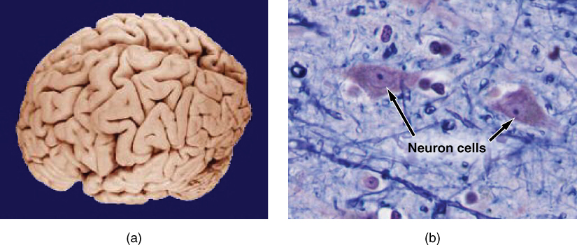
Like most scientific disciplines, anatomy has areas of specialization. Gross anatomy is the study of the larger structures of the body, those visible without the aid of magnification (Figure 1.2 a). Macro- means “large,” thus, gross anatomy is also referred to as macroscopic anatomy. In contrast, micro- means “small,” and microscopic anatomy is the study of structures that can be observed only with the use of a microscope or other magnification devices (Figure 1.2 b). Microscopic anatomy includes cytology, the study of cells and hishistologytology, the study of tissues. As the technology of microscopes has advanced, anatomists have been able to observe smaller and smaller structures of the body, from slices of large structures like the heart, to the three-dimensional structures of large molecules in the body.
Figure 1.2 Gross and Microscopic Anatomy (a) Gross anatomy considers large structures such as the brain. (b) Microscopic anatomy can deal with the same structures, though at a different scale. This is a micrograph of nerve cells from the brain. LM × 1600. (credit a: “WriterHound”/Wikimedia Commons; credit b: Micrograph provided by the Regents of University of Michigan Medical School © 2012)
Anatomists take two general approaches to the study of the body’s structures: regional and systemic. Regional anatomy is the study of the interrelationships of all of the structures in a specific body region, such as the abdomen. Studying regional anatomy helps us appreciate the interrelationships of body structures, such as how muscles, nerves, blood vessels, and other structures work together to serve a particular body region. In contrast, systemic anatomy is the study of the structures that make up a discrete body system—that is, a group of structures that work together to perform a unique body function. For example, a systemic anatomical study of the muscular system would consider all of the skeletal muscles of the body.
Whereas anatomy is about structure, physiology is about function. Human physiology is the scientific study of the chemistry and physics of the structures of the body and the ways in which they work together to support the functions of life. Much of the study of physiology centers on the body’s tendency toward homeostasis. Homeostasis is the state of steady internal conditions maintained by living things. The study of physiology certainly includes observation, both with the naked eye and with microscopes, as well as manipulations and measurements. However, current advances in physiology usually depend on carefully designed laboratory experiments that reveal the functions of the many structures and chemical compounds that make up the human body.
Like anatomists, physiologists typically specialize in a particular branch of physiology. For example, neurophysiology is the study of the brain, spinal cord, and nerves and how these work together to perform functions as complex and diverse as vision, movement, and thinking. Physiologists may work from the organ level (exploring, for example, what different parts of the brain do) to the molecular level (such as exploring how an electrochemical signal travels along nerves).
Form is closely related to function in all living things. For example, the thin flap of your eyelid can snap down to clear away dust particles and almost instantaneously slide back up to allow you to see again. At the microscopic level, the arrangement and function of the nerves and muscles that serve the eyelid allow for its quick action and retreat. At a smaller level of analysis, the function of these nerves and muscles likewise relies on the interactions of specific molecules and ions. Even the three-dimensional structure of certain molecules is essential to their function.
Your study of anatomy and physiology will make more sense if you continually relate the form of the structures you are studying to their function. In fact, it can be somewhat frustrating to attempt to study anatomy without an understanding of the physiology that a body structure supports. Imagine, for example, trying to appreciate the unique arrangement of the bones of the human hand if you had no conception of the function of the hand. Fortunately, your understanding of how the human hand manipulates tools—from pens to cell phones—helps you appreciate the unique alignment of the thumb in opposition to the four fingers, making your hand a structure that allows you to pinch and grasp objects and type text messages.
Structural Organization
Learning Objectives
By the end of this section, you will be able to:
- Describe the structure of the human body in terms of six levels of organization in order from simplest to most complex.
- List the eleven organ systems of the human body and identify at least one organ and one major function of each
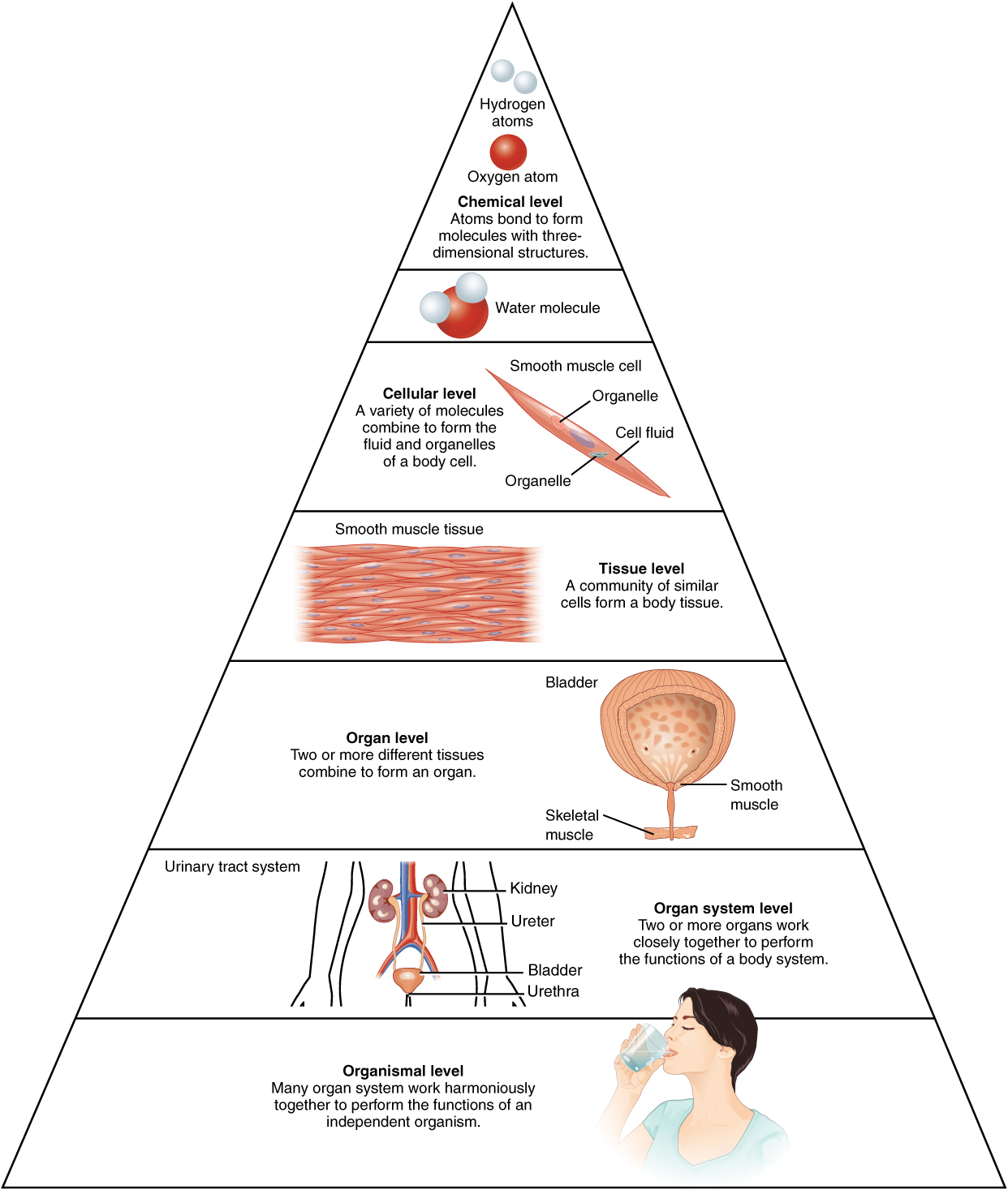
Before you begin to study the different structures and functions of the human body, it is helpful to consider its basic architecture; that is, how its smallest parts are assembled into larger structures. It is convenient to consider the structures of the body in terms of fundamental levels of organization that increase in complexity: subatomic particles, atoms, molecules, organelles, cells, tissues, organs, organ systems, organisms and biosphere (Figure 1.3).
Figure 1.3 Levels of Structural Organization of the Human Body The organization of the body often is discussed in terms of six distinct levels of increasing complexity, from the smallest chemical building blocks to a unique human organism.
The Levels of Organization
To study the chemical level of organization, scientists consider the simplest building blocks of matter: subatomic particles, atoms and molecules. All matter in the universe is composed of one or more unique pure substances called elements, familiar examples of which are hydrogen, oxygen, carbon, nitrogen, calcium, and iron. The smallest unit of any of these pure substances (elements) is an atom. Atoms are made up of subatomic particles such as the proton, electron and neutron. Two or more atoms combine to form a molecule, such as the water molecules, proteins, and sugars found in living things. Molecules are the chemical building blocks of all body structures.
A cell is the smallest independently functioning unit of a living organism. Even bacteria, which are extremely small, independently-living organisms, have a cellular structure. Each bacterium is a single cell. All living structures of human anatomy contain cells, and almost all functions of human physiology are performed in cells or are initiated by cells.
A human cell typically consists of flexible membranes that enclose cytoplasm, a water-based cellular fluid together with a variety of tiny functioning units called organelles. In humans, as in all organisms, cells perform all functions of life. A tissue is a group of many similar cells (though sometimes composed of a few related types) that work together to perform a specific function. An organ is an anatomically distinct structure of the body composed of two or more tissue types. Each organ performs one or more specific physiological functions. An organ system is a group of organs that work together to perform major functions or meet physiological needs of the body.
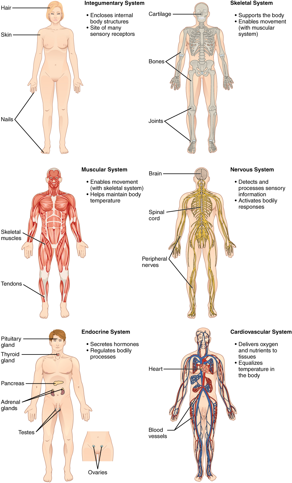
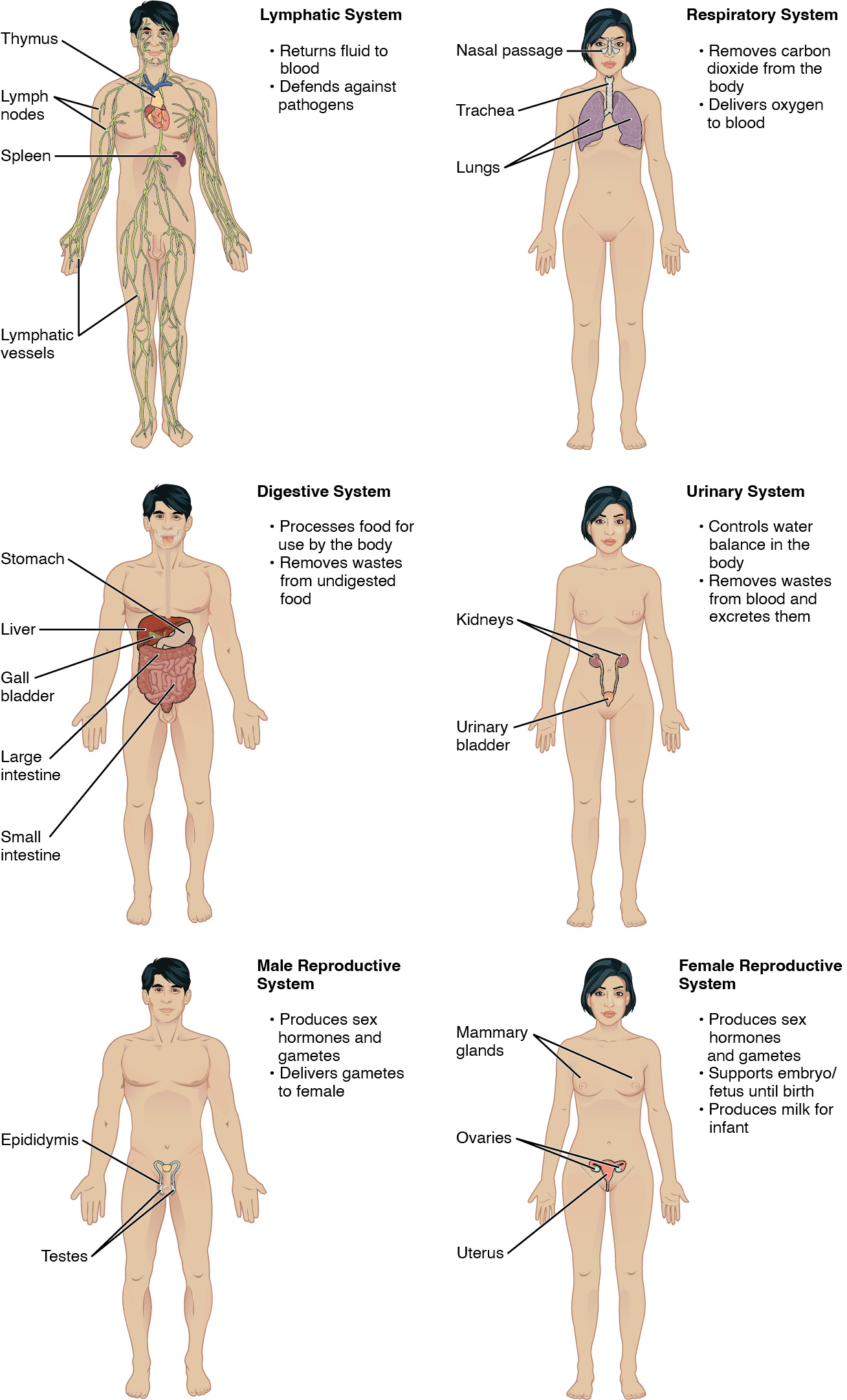
This book covers eleven distinct organ systems in the human body (Figure 1.4 and Figure 1.5). Assigning organs to organ systems can be imprecise since organs that “belong” to one system can also have functions integral to another system. In fact, most organs contribute to more than one system.
In this book and throughout your studies of biological sciences, you will often read descriptions related to similarities and differences among biological structures, processes, and health related to a person’s biological sex. People often use the words “female” and “male” to describe two different concepts: our sense of gender identity, and our biological sex as determined by our chromosomes, hormones, organs, and other physical characteristics. For some people, gender identity is different from biological sex or their sex assigned at birth. Throughout this book, “female” and “male” refer to sex only, and the typical anatomy and physiology of XX and XY individuals is discussed.
Figure 1.4 Organ Systems of the Human Body Organs that work together are grouped into organ systems.
Figure 1.5 Organ Systems of the Human Body (continued) Organs that work together are grouped into organ systems.
The organism level is the highest level or most complex level of organization for the purpose of studying human biology. An organism is a living being that has a cellular structure and that can independently perform all physiologic functions necessary for life. In multicellular organisms, including humans, all cells, tissues, organs, and organ systems of the body work together to maintain the life and health of the organism.
Homeostasis
Learning Objectives
By the end of this section, you will be able to:
- Discuss the role of homeostasis in healthy functioning
- Contrast negative and positive feedback, giving one physiologic example of each mechanism
Homeostasis describes how a cell or organism senses and responds to its environment in order to maintain a stable internal state. Maintaining homeostasis requires that the body continuously monitor its internal conditions. From body temperature to blood pressure to levels of certain nutrients, each physiological condition has a particular set point.
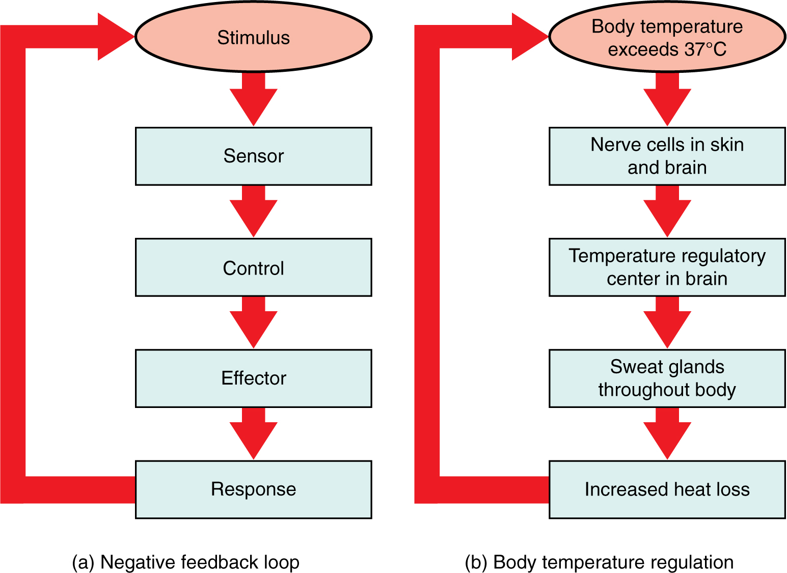
There are three components that interact to maintain homeostasis. (Figure 1.10a). A receptor, also referred to a receptor, is a component of a feedback system that monitors a stimulus or physiological value. This value is reported to the control center. The control center is the component in a feedback system that compares the value to the normal range and determines if a response is required. If the value deviates too much from the set point, then the control center activates an effector. An effector i is the component in a feedback system that carries out the response. The most common type of effector is muscle and glands. Muscles will contract and glands will secrete.
A set point is the physiological value around which the normal range fluctuates. A normal range is the restricted set of values that is optimally healthful and stable. For example, the set point for normal human body temperature is approximately 37°C (98.6°F). Physiological parameters, such as body temperature and blood pressure, tend to fluctuate within a normal range a few degrees above and below that point. Control centers in the brain and other parts of the body monitor and react to deviations from homeostasis maintain normal body temperature. Keep in mind, that the set-point may be adjusted or modified.
Negative Feedback
Negative feedback is the most common type of homeostatic control mechanism. The purpose of negative feedback is to maintain a set point by removing the stimulus. Therefore, negative feedback maintains body parameters within their normal range. The maintenance of homeostasis by negative feedback goes on throughout the body at all times, and an understanding of negative feedback is thus fundamental to an understanding of human physiology.
Figure 1.10 Negative Feedback System In a negative feedback system, a stimulus—a deviation from a set point—is resisted through a physiological process that returns the body to homeostasis. (a) A negative feedback system has five basic parts. (b) Body temperature is regulated by negative feedback.
In order to set the system in motion, a stimulus must drive a physiological parameter beyond its normal range (that is, beyond homeostasis). This stimulus is detected by a specific sensor. For example, in the control of blood glucose, specific endocrine cells in the pancreas detect excess glucose (the stimulus) in the bloodstream. These pancreatic beta cells respond to the increased level of blood glucose by releasing the hormone insulin into the bloodstream. The insulin signals skeletal muscle fibers, fat cells (adipocytes), and liver cells to take up the excess glucose, removing it from the bloodstream. As glucose concentration in the bloodstream drops, the decrease in concentration—the actual negative feedback—is detected by pancreatic alpha cells, and insulin release stops. This prevents blood sugar levels from continuing to drop below the normal range.
Humans have a similar temperature regulation feedback system that works by promoting either heat loss or heat gain (Figure 1.10b). When the brain’s temperature regulation center receives data from the sensors indicating that the body’s temperature exceeds its normal range, it stimulates a cluster of brain cells referred to as the “heat-loss center.” This stimulation has three major effects:
- Blood vessels in the skin begin to dilate allowing more blood from the body core to flow to the surface of the skin allowing the heat to radiate into the environment.
- As blood flow to the skin increases, sweat glands are activated to increase their output. As the sweat evaporates from the skin surface into the surrounding air, it takes heat with it.
- The depth of respiration increases, and a person may breathe through an open mouth instead of through the nasal passageways. This further increases heat loss from the lungs.
In contrast, activation of the brain’s heat-gain center by exposure to cold reduces blood flow to the skin, and blood returning from the limbs is diverted into a network of deep veins. This arrangement traps heat closer to the body core and restricts heat loss. If heat loss is severe, the brain triggers an increase in random signals to skeletal muscles, causing them to contract and producing shivering. The muscle contractions of shivering release heat while using up ATP. The brain triggers the thyroid gland in the endocrine system to release thyroid hormone, which increases metabolic activity and heat production in cells throughout the body. The brain also signals the adrenal glands to release epinephrine (adrenaline), a hormone that causes the breakdown of glycogen into glucose, which can be used as an energy source. The breakdown of glycogen into glucose also results in increased metabolism and heat production.
Interactive Link
Water concentration in the body is critical for proper functioning. A person’s body retains very tight control on water levels without conscious control by the person. Watch this video to learn more about water concentration in the body. Which organ has primary control over the amount of water in the body?
Positive Feedback
Positive feedback intensifies a change in the body’s physiological condition rather than reversing it. A deviation from the normal range results in more change, and the system moves farther away from the normal range or increases the stimulus. Positive feedback in the body is normal only when there is a definite end point. Childbirth and the body’s response to blood loss are two examples of positive feedback loops that are normal but are activated only when needed.
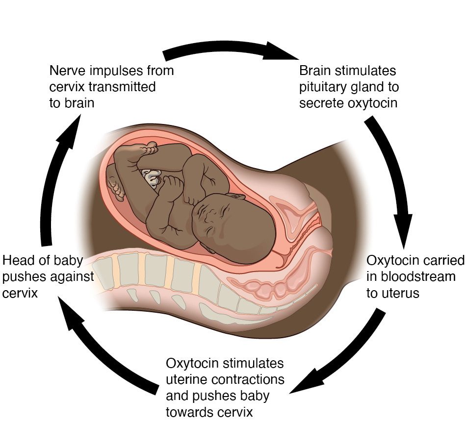
Childbirth at full term is an example of a situation in which the maintenance of the existing body state is not desired. Enormous changes in a person’s body are required to expel the baby at the end of pregnancy. And the events of childbirth, once begun, must progress rapidly to a conclusion or the life of a person giving birth and the baby are at risk. The extreme muscular work of labor and delivery are the result of a positive feedback system (Figure 1.11).
The first contractions of labor push the baby toward the cervix (the lowest part of the uterus); the pressure on the cervix is the stimulus. The cervix contains stretch-sensitive nerve cells that monitor the degree of stretching (the sensors). These nerve cells (pressure receptors/sensors) send messages to the brain, which in turn causes the pituitary gland at the base of the brain to release the hormone oxytocin into the bloodstream. Oxytocin causes stronger contractions of the smooth muscles in of the uterus (the effectors), pushing the baby further down the birth canal. This causes even greater stretching and pressure on the cervix. The cycle of stretching, oxytocin release, and increasingly more forceful contractions stops only when the baby is born. At this point, the stretching and pressure of the cervix halts, stopping the release of oxytocin.
Figure 1.11 Positive Feedback Loop Normal childbirth is driven by a positive feedback loop. A positive feedback loop results in a change in the body’s status, rather than a return to homeostasis.
A second example of positive feedback centers on reversing extreme damage to the body. Following a penetrating wound, the most immediate threat is excessive blood loss. Less blood circulating means reduced blood pressure and reduced perfusion (penetration of blood) to the brain and other vital organs. If perfusion is severely reduced, vital organs will shut down and the person will die. The body responds to this potential catastrophe by releasing substances in the injured blood vessel wall that begin the process of blood clotting. As each step of clotting occurs, it stimulates the release of more clotting substances. This accelerates the processes of clotting and sealing off the damaged area. Clotting is contained in a local area based on the tightly controlled availability of clotting proteins. This is an adaptive, life-saving cascade of events.
The study of structure of living things. Anatomy includes composition (what its made of--molecules, cells, tissues, etc.) and location in the body.
Study of anatomy that can be seen with the naked eye (macroscopic).
The study of cells and tissues (microscopic).
The study of the function of living things, it includes chemical reactions that carry out the functions.
Describes how a cell or organism can sense and respond to its environment in order to maintain a stable internal state.
The smallest unit of an element that can enter into a chemical reaction.
Two or more atoms held together by chemical bonds.
The smallest unit of life.
A group of similar cells that work together to perform a specific function(s).
Two or more tissues that work together to perform a specific function(s).
Two or more organs that work together to perform a specific function(s).
An entire living being.
Specialized cell that monitors a change in a stimulus.
Compares incoming sensory information to a set-point and determines if a response is necessary.
A muscle or gland that carries out a response. A muscle will contract and a gland will secrete.
Physiological value around which the normal range fluctuates.
Feedback mechanism that works to increase the stimulus.

